Opening a Window to an Unseen World
Researchers in the Agricultural Research Service (ARS) Electron and Confocal Microscopy Unit can magnify a cell’s internal structures to 200,000 times their size, flash freeze mites in liquid nitrogen to create striking “snapshots” as they feed, and create color-enhanced images that show a virus infecting its host. The resulting images help scientists determine how agricultural pests and pathogens feed, reproduce, respond to threats, and survive.

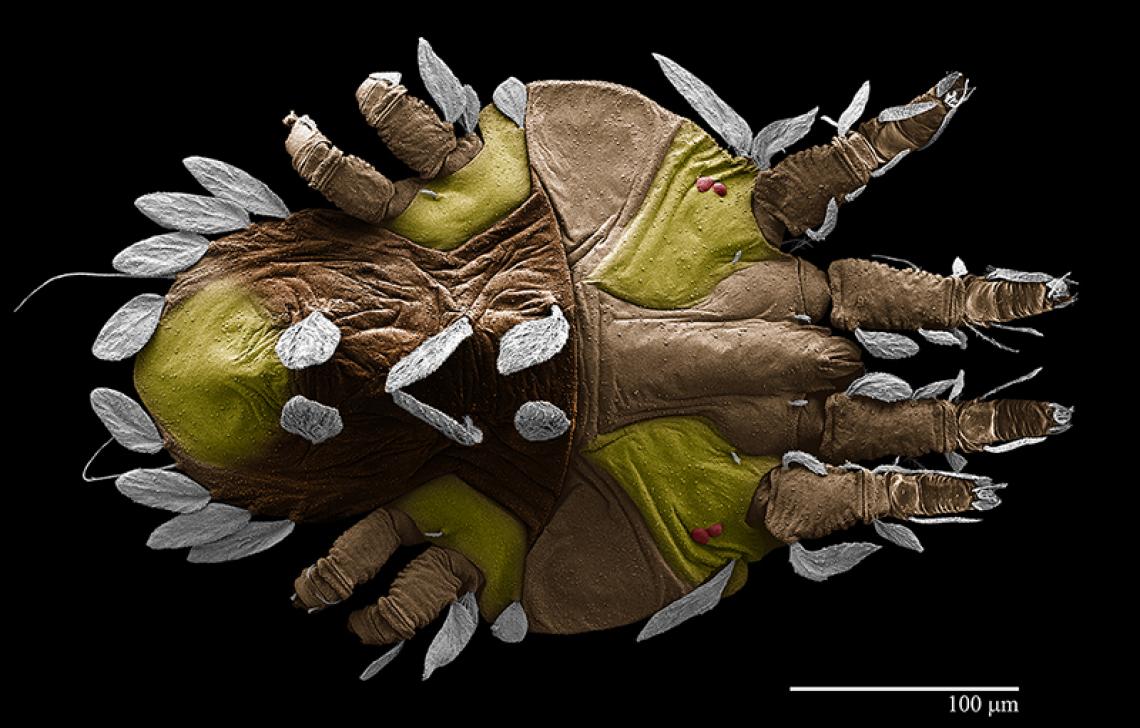
Images like this one – of a flat mite that feeds on coffee plants – have won scientific photography awards for the Electron and Confocal Microscopy Unit, graced the covers of prestigious scientific journals, and enhanced our scientific understanding of many of the microbes, pests, and pathogens that attack crops, infect livestock, and make people sick. (D4049-1)
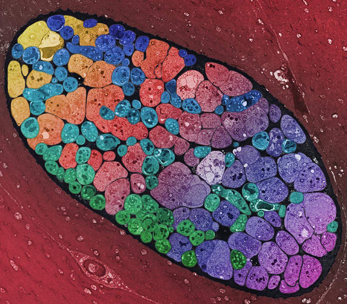
A sampling of the unit’s digital photo album shows the eclectic nature of its efforts.
The team also has a unique 3D printing capability that allows them to transform the images they create into hand-size 3D models that are the most structurally accurate models of mites and other organisms currently available. The researchers hope that one day they will be able to upload the 3D files to an online database so that anyone with a 3D printer can reproduce them to use as instructional aids, in research, or for scientific outreach.


